
Safranin O
Description
Safranin O is a cationic (basic) biological stain commonly used in histology and microbiology for its affinity for acidic cellular components, particularly nucleic acids and acidic polysaccharides. It appears as a red or pink dye and is frequently employed as a counterstain in techniques such as Gram staining, where it stains Gram-negative bacteria red after the primary crystal violet stain is washed out. In plant histology, Safranin O selectively stains lignified, suberized, or cutinized cell walls, xylem, and nuclei, providing clear contrast against other tissues when used with fast green or other counterstains. Its staining mechanism relies on electrostatic interactions between the positively charged dye and negatively charged cellular structures, making it highly useful for highlighting structural details in both microbial and plant specimens.
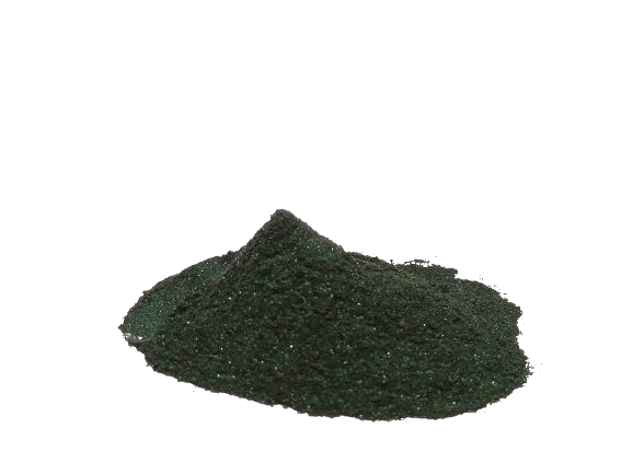
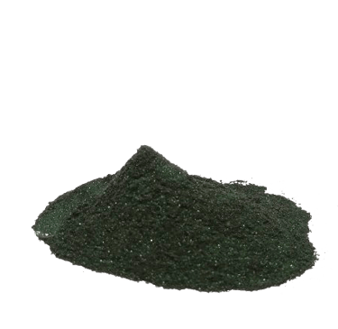
Appearance of Safranin O
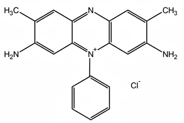
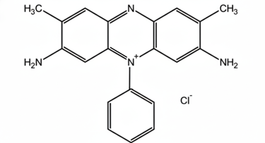
Structure of Safranin O
Dye Content
(Spectrophotometry)
Solubility
Appearance
Molecular Weight
Synonyms
IUPAC Name
CI No.
CAS No.
Molecular Formula
C₁₀H₁₀ClN₃
477-73-6
50240
3,7-diamino-5- phenylphenazinium chloride
BASIC RED-2
350.85 g/mol
reddish-orange crystalline powder or red solution.
Soluble in water
98%
Uses
1. Microbiology Uses
Gram Staining:
Safranin O is the counterstain in the Gram staining procedure. After Gram-positive bacteria retain the crystal violet-iodine complex, Gram-negative bacteria are decolorized with alcohol and then take up Safranin O, appearing red or pink. This contrast allows differentiation between Gram-positive (purple) and Gram-negative (red) bacteria under a microscope.Capsule and Spore Visualization:
Though not the primary stain, Safranin O can sometimes be used to provide background staining in spore staining techniques, making spores (which remain unstained) easier to visualize.Bacterial Morphology Studies:
Safranin O stains bacterial cytoplasm lightly, allowing observation of cell shape, arrangement, and size.
2. Histology Uses
Plant Tissue Staining:
Safranin O is heavily used in plant histology because it stains lignified, suberized, or cutinized cell walls red. Examples:Xylem vessels: Highlighting woody tissue.
Sclerenchyma cells: Secondary cell walls stand out.
Nuclei: Provides contrast in tissues when combined with other stains like fast green (which stains cellulose and cytoplasm green).
Combination Stains:
Safranin-Fast Green: Common for plant tissue sections. Safranin stains lignified or suberized structures red, and fast green stains cytoplasm or non-lignified tissue green.
Double Staining: In bone and cartilage studies, Safranin O can stain proteoglycans in cartilage red, allowing distinction from surrounding bone tissue.
3. Cartilage and Connective Tissue Studies
Glycosaminoglycan Detection:
Safranin O has a high affinity for sulfated glycosaminoglycans in cartilage and connective tissue, making it an excellent stain for evaluating cartilage integrity in osteoarthritis research.Histopathology Applications:
It is used to assess cartilage degeneration, repair, and matrix composition by quantitatively measuring staining intensity.
4. Cytology and General Uses
Nuclear Staining:
Safranin O binds to nucleic acids, staining nuclei red in plant and some animal tissues.Cytoplasmic Background Staining:
Provides contrast against basophilic stains in cytological preparations, helping identify cellular components.
5. Special Applications
Biomaterial and Tissue Engineering Research:
Used to visualize cartilage scaffolds and extracellular matrix components in tissue engineering.Forensic Botany and Paleobotany:
Highlights lignified or preserved plant structures in ancient samples.Medical Diagnosis:
Helps detect cartilage and connective tissue abnormalities in biopsies.
Export Worthy packing.
Storage Conditions
Store in room temperature.
Packing
:
:
:
:
:
:
:
:
:
Contact
Reach out with questions or service requests
Phone
© 2023-2025, All Rights reserved by AADHAV Remedies Pvt Ltd.