
Basic Yellow-1
Description
Thioflavin T is a bright yellow crystalline powder widely used as a fluorescent dye in biochemical and biomedical research, particularly for detecting amyloid fibrils associated with protein misfolding and neurodegenerative diseases. Chemically, it is a benzothiazolium salt with the molecular formula C₁₇H₁₉ClN₂S and a molecular weight of approximately 318.86 g/mol. It is moderately soluble in water and soluble in organic solvents like methanol. When bound to amyloid fibrils, Thioflavin T exhibits a characteristic shift in its fluorescence emission (~482 nm), enabling sensitive visualization of beta-sheet aggregates under microscopy. Its applications extend to laboratory staining, aggregation assays, and research on diseases such as Alzheimer’s. The compound should be handled with care, as it can irritate skin, eyes, and the respiratory tract, and it is typically sold for research use only.
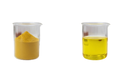
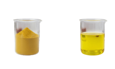
Appearance of Basic Yellow -1
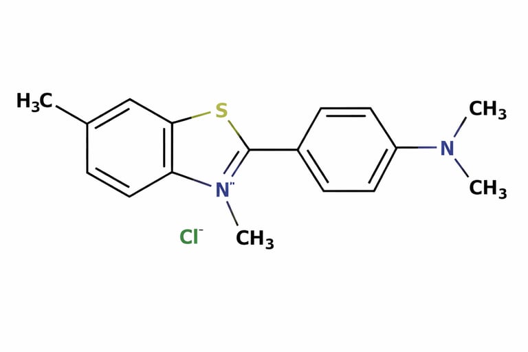
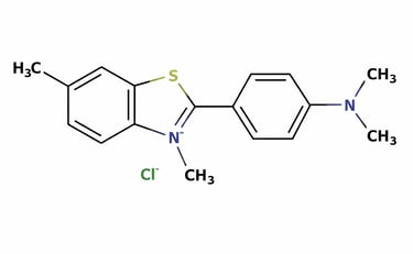
Structure of Basic Yellow -1
Dye Content (Spectrophotometry)
Solubility
Appearance
Molecular Weight
Synonyms
IUPAC Name
CI No.
CAS No.
Molecular Formula
C₁₇H₁₉ClN₂S
2390‑54‑7
50220
2‑[4‑(dimethy lamino)phenyl]‑3, 6‑dimethyl‑1, 3‑benzothiazol‑ 3‑ium chloride
Thioflavin T
318.86 g/mol
Bright yellow crystalline powder
Soluble in water
98%
Uses
1. Biomedical Research and Diagnostics
Amyloid Detection: Thioflavin T is primarily used as a fluorescent probe to detect amyloid fibrils, which are protein aggregates with beta-sheet structures. These fibrils are associated with neurodegenerative disorders such as Alzheimer’s disease, Parkinson’s disease, and Huntington’s disease.
Fluorescence Assays: Upon binding to amyloid fibrils, Thioflavin T exhibits a shift in its fluorescence spectrum (excitation ~450 nm; emission ~482 nm). This allows researchers to quantify fibril formation in vitro or track aggregation kinetics over time.
Histopathology: Used in tissue staining to visualize amyloid plaques in biopsy samples or post-mortem brain sections. Under fluorescence microscopy, amyloid deposits fluoresce bright yellow-green, distinguishing them from other tissue components.
High-throughput Screening: Thioflavin T is incorporated into plate-based assays to screen for compounds that inhibit or promote amyloid fibril formation, making it critical in drug discovery for neurodegenerative diseases.
2. Protein Aggregation Studies
Mechanistic Studies: Thioflavin T helps researchers study protein misfolding pathways by providing a fluorescent readout of fibril nucleation and elongation.
Kinetics and Thermodynamics: It enables real-time monitoring of aggregation kinetics, measuring lag phase, growth rate, and saturation of fibril formation.
Comparative Analysis: Different protein mutants or environmental conditions (pH, temperature, ionic strength) can be compared for their propensity to form fibrils using Thioflavin T fluorescence intensity as a metric.
3. Laboratory Research and Biochemistry
Fluorescent Staining: Beyond amyloid detection, Thioflavin T can be used to stain amyloid-like structures in vitro, such as synthetic fibrils formed by peptides or proteins.
Spectroscopic Analysis: Researchers use Thioflavin T to study conformational changes in proteins, as its binding is sensitive to the presence of beta-sheet secondary structure.
Microscopy and Imaging: Compatible with fluorescence microscopy, confocal microscopy, and high-resolution imaging, it provides precise localization of fibrillar aggregates within cells or tissue samples.
4. Research Tool for Neurodegenerative Disease Models
Alzheimer’s Disease Models: Thioflavin T is used to visualize amyloid-beta plaques in animal models or cultured neuronal cells.
Parkinson’s Disease Models: Can be applied to detect alpha-synuclein fibrils in experimental setups.
Therapeutic Research: Facilitates testing of anti-aggregation compounds or small molecules that may prevent fibril formation or promote fibril disassembly.
5. Chemical and Physical Characterization
Spectroscopy Studies: Its interaction with fibrils is used in UV-Vis, fluorescence spectroscopy, and circular dichroism to analyze fibril formation.
Structure-Function Correlation: Thioflavin T helps correlate structural changes in proteins with aggregation propensity and toxicity.
Quantitative Assays: Fluorescence intensity is often proportional to fibril quantity, allowing for semi-quantitative or quantitative analysis.
Packing
:
Storage Conditions
Store in room temperature.
6.Educational and Demonstration Uses
Teaching Tool: In biochemistry or cell biology labs, Thioflavin T can be used to demonstrate protein aggregation visually.
Visualization Experiments: Its fluorescent properties make it ideal for interactive demonstrations of amyloid detection techniques.
Export Worthy packing.
:
:
:
:
:
:
:
:
Contact
Reach out with questions or service requests
Phone
© 2023-2025, All Rights reserved by AADHAV Remedies Pvt Ltd.